Tooth preservation depends on pocket depth, bone loss, furcation and mobility. F3 teeth with bone loss generally warrant extraction, while flap surgery can help selected cases.
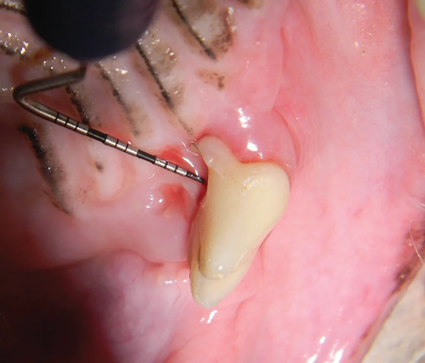
Choosing whether to preserve a dog’s periodontally diseased tooth depends on what examination and imaging reveal, and on whether the owner can sustain home care. Pocket depth, loss of bone attachment, furcation involvement and tooth mobility all inform the decision. Veterinarians assess these findings under anesthesia, using periodontal probing and intraoral dental radiographs, with cone beam computed tomography available when needed. Each measure describes a different part of the tooth’s support, so no single finding should be read in isolation. The goal is to judge whether the tooth can be treated and then maintained, not simply whether it remains in place today.
- Stage 1 (F1): Attachment loss extends less than halfway beneath the crown of a multirooted tooth, in any direction.
- Stage 2 (F2): Attachment loss extends more than halfway beneath the crown, but does not pass through the furcation.
- Stage 3 (F3): Attachment loss passes through the furcation, from one side beneath the crown to the other.
- Stage 0 (M0): Physiologic movement of up to 0.2 mm.
- Stage 1 (M1): Increased movement in a nonaxial direction, between 0.2 mm and 0.5 mm.
- Stage 2 (M2): Increased movement in a nonaxial direction, between 0.5 mm and 1 mm.
- Stage 3 (M3): Nonaxial movement greater than 1 mm, or any axial movement.
These measurements need context. Healthy mandibular incisors, for example, may show apparent bone loss and mobility while retaining normal gingival attachment; in dogs, a normal sulcus is less than 3 mm deep.2-4 That distinction matters because a measurement that looks alarming on its own may not establish disease. Clinicians need to compare what they feel during probing with what the radiographs show and where the tooth sits in the mouth. The staging systems provide a shared way to describe damage, but they don’t make the treatment decision by themselves.
When flap surgery is needed
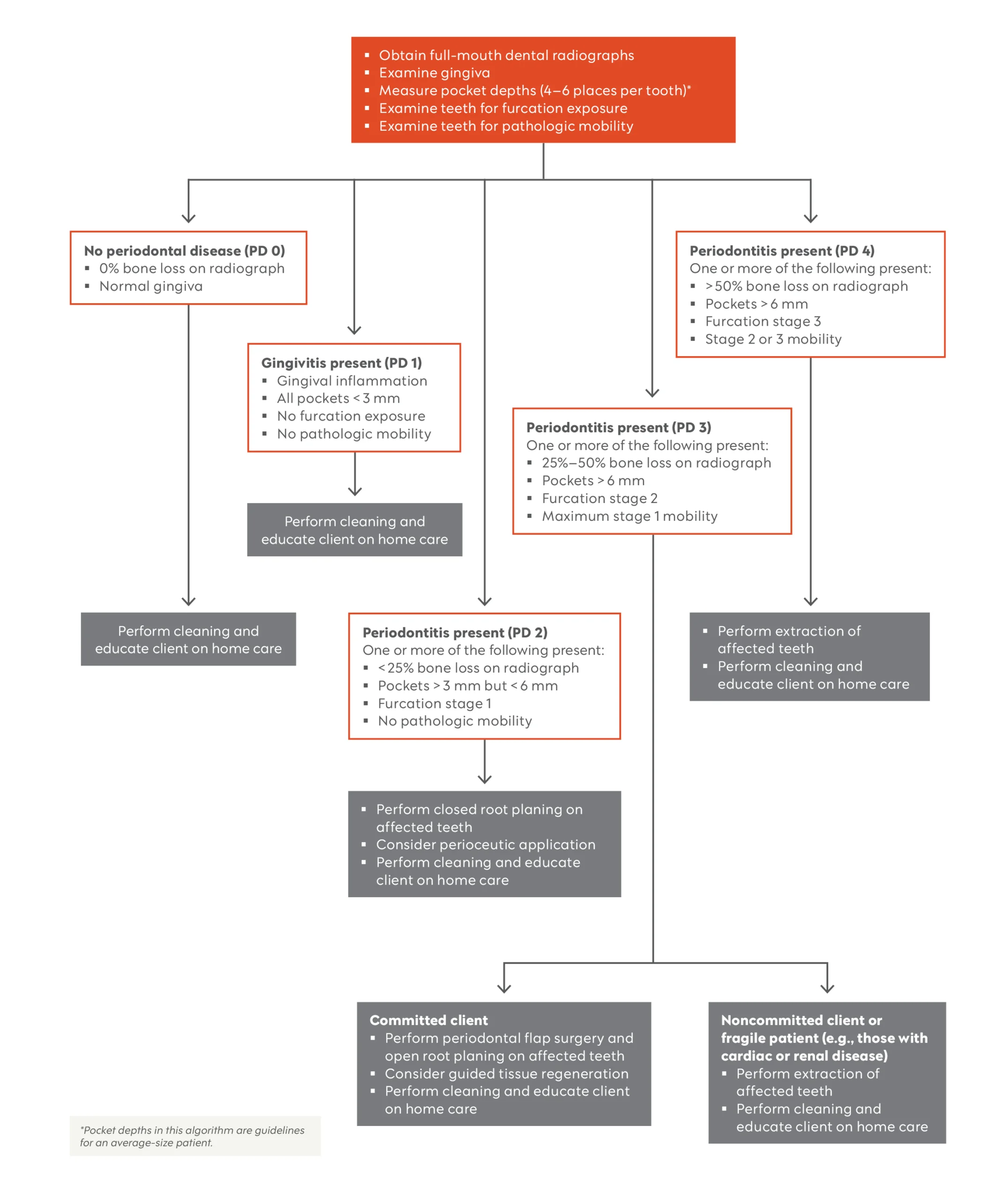
Furcation stage and pocket depth help determine whether a periodontal flap is required. F1 teeth can be treated with closed cleaning, but F2 and F3 furcations need a flap to expose the root surfaces. Pockets deeper than 6 mm also call for flap surgery.4-10 The practical issue is access: treatment has to reach the surfaces where disease is present. A tooth’s stage therefore affects not only how much support has been lost, but also whether cleaning can be effective without surgery.
Without direct access, plaque, calculus and granulation tissue can remain below the gumline. The disease may then progress between professional cleanings, since owners can’t effectively clean those concealed surfaces at home. This is a limitation of the treatment plan, not a failure of diligence: brushing cannot reach every surface hidden in a deep pocket or furcation. Surgery can expose those areas for cleaning, but it also creates a continuing obligation to maintain the result. If that ongoing care isn’t realistic, the expected benefit of preserving the tooth changes.
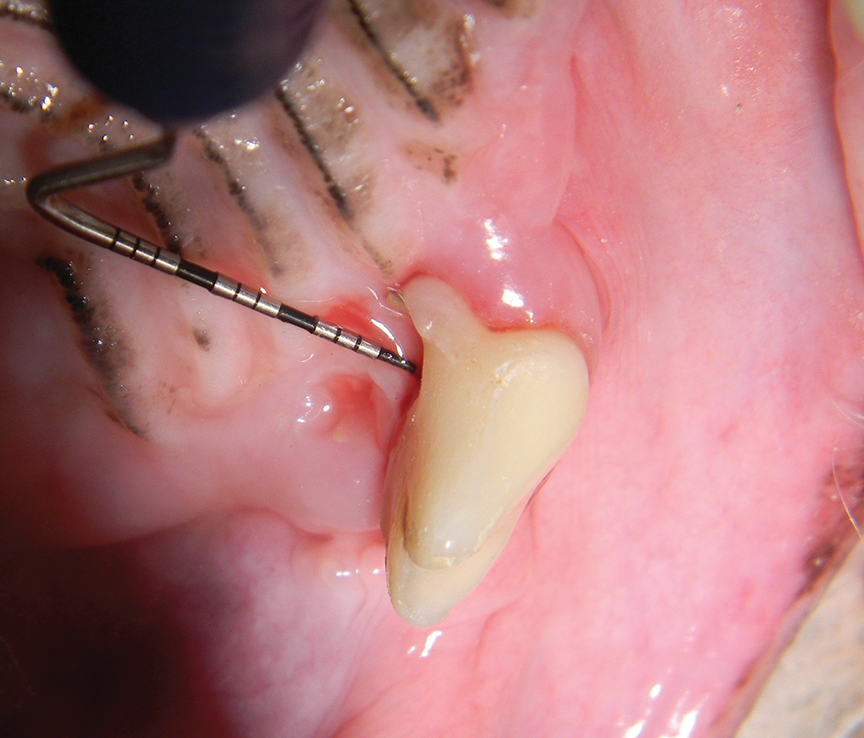
Figure 1. A maxillary fourth premolar with stage 2 furcation. Effective cleaning requires a periodontal flap. With flap surgery and guided tissue regeneration, the tooth has a very good prognosis; if referral isn’t available, extraction is recommended. The example shows why a stage alone doesn’t settle the choice: access to treatment and the prospects for follow-up also matter.
When extraction is the sounder choice
Some teeth can’t be predictably maintained, even with surgery and diligent home care. The article’s author recommends extracting all F3 teeth with bone loss. In these cases, persistent periodontal inflammation can continue to advance; horizontal bone loss in an F3 tooth can’t reliably be repaired with guided tissue regeneration. Bone can regrow laterally in infrabony pockets and F2 teeth, but not vertically. The distinction is central to the recommendation: a procedure’s potential to improve a defect depends on its shape and location, as well as the amount of disease.
An F3 tooth may look relatively healthy and have little bone loss, yet its through-and-through furcation remains a concern. The author cites the focal infection theory, under which periodontal inflammation has local and systemic effects, as a reason not to treat appearance alone as evidence that the tooth can be preserved.11-15 A reassuring view of the crown can’t reveal the full condition of the supporting structures. This is more significant than it looks: decisions based on appearance risk overlooking disease in the areas that are hardest to inspect without probing and imaging.
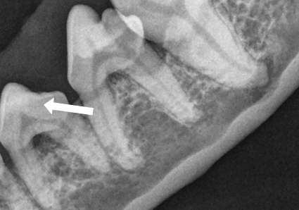
Figure 2. Premolar 307 has stage 3 furcation involvement, extending through the furcation (arrow). The recommendation is extraction, despite minimal bone loss. Here, the furcation’s through-and-through involvement is the key finding, even though the amount of visible bone loss is limited.
Patient health and owner capacity also shape the plan. If an owner can’t provide home care, or comorbidities make future anesthesia unsuitable, extraction should be recommended. For severe disease, safety concerns or a preference to pursue tooth preservation, referral to a board-certified veterinary dentist is advised. The treatment choice has to account for what can be done now and what the patient is likely to tolerate later. A technically possible preservation procedure isn’t automatically a practical one.
Plan for ongoing maintenance
Most patients should receive professional dental cleaning under anesthesia annually, even when the gums look healthy.16 Smaller-breed dogs, dogs and cats that are more susceptible to plaque and periodontal disease, and animals that have undergone advanced flap procedures may need more frequent cleanings. The interval should reflect the individual patient’s risks and treatment history, rather than a calendar alone. For teeth that are preserved, that maintenance—and effective home care—helps determine whether treatment can hold. A successful procedure is only one part of the outcome.
What the decision means for owners
The framework asks owners and veterinarians to weigh three connected questions: how much support the tooth has lost, whether diseased surfaces can be reached and treated, and whether follow-up care is feasible. A favorable answer to only one question may not be enough. That said, the staging details help make the trade-offs clearer and give owners a basis for discussing why a tooth that appears usable may still be a poor candidate for preservation.
If you’re working through this decision with a veterinarian, ask what the probing and imaging show, what treatment would be required, and what home care would look like afterward. The answer may differ between teeth in the same mouth, because their pocket depth, mobility and furcation involvement can differ. Here’s the thing: preservation is a continuing plan, not a one-time procedure. And yet, the article’s examples also show that some teeth can have a very good prognosis when treatment gives clinicians adequate access and maintenance can continue. The details matter.
The post Periodontal Disease in Dogs: Staging and Treatment Algorithm appeared first on Today's Veterinary Practice.
Discussion
Sign in to join the discussion.