Sublingual swelling in cats can resemble squamous cell carcinoma but may have other causes, including ranulas and eosinophilic granulomas.
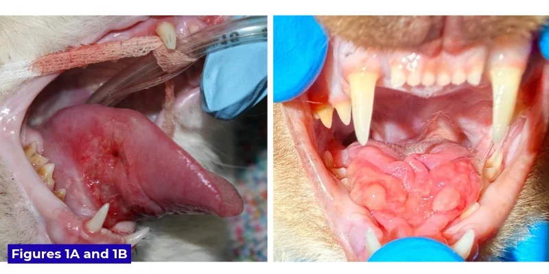
Squamous cell carcinoma (SCC) is a leading concern when an older cat develops bloody drool, reduced appetite and difficulty swallowing. But sublingual swelling has other causes, and the visual resemblance between them makes tissue diagnosis important. Ranulas, eosinophilic granulomas and foreign bodies belong on the differential list.
Ranulas: saliva collecting beneath the tongue
A ranula is a smooth swelling under the tongue caused by saliva accumulating after damage to a salivary gland or duct. The name comes from the Latin rana, or frog, because the swelling can resemble a frog’s underside. The mandibular and sublingual ducts open at paired structures called caruncles near the back of the lower jaw’s midline. Damage to the caruncles or the ducts that connect them to the glands can allow saliva to leak into surrounding tissue.
Ranulas may affect one or both sides and are usually not inflamed. If they become large, however, contact with the teeth can cause trauma. Some resolve without intervention; others require surgery to remove the mandibular and sublingual glands.
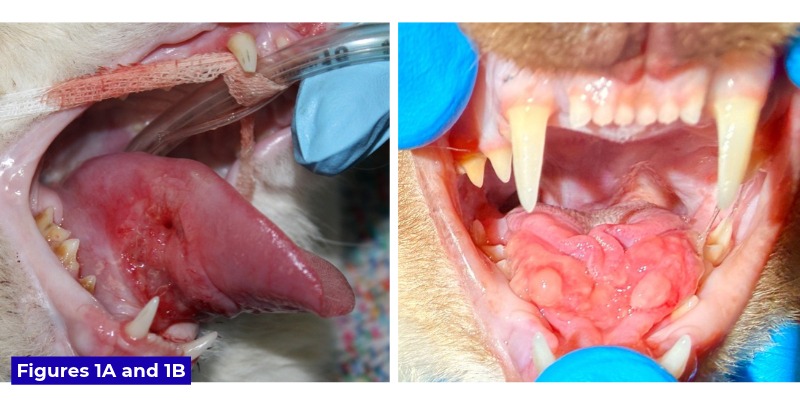
Postoperative swelling can also resemble a ranula. When oral tumor surgery requires salivary ducts to be cut and ligated during a mandibulectomy, temporary sublingual edema may follow. These pseudoranulas typically subside within a few days. A 1978 study found no ranulas in dogs after experimental injury to a gland or duct, though the study involved few animals. The author has also seen a ranula develop after duct transection during cancer surgery.
Eosinophilic disease can mimic both SCC and ranulas
Upper-lip ulcers, often called rodent ulcers, are a familiar sign of eosinophilic granuloma in cats. The condition can also cause non-ulcerated swelling of the lips or the tissue beneath the tongue. A sublingual lesion may be smooth like a ranula or firm and ulcerated like SCC, so appearance alone may not settle the diagnosis.
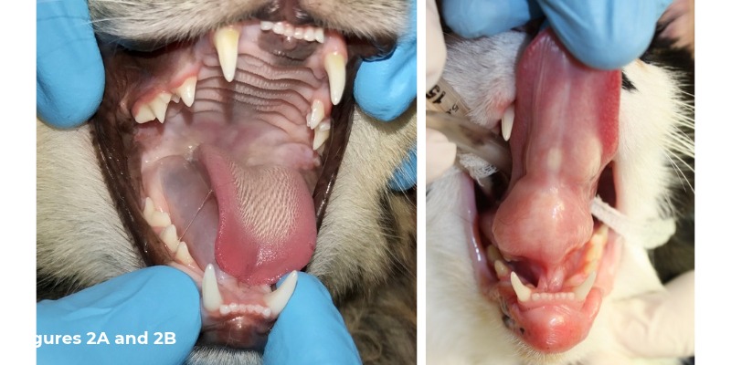
Milo, a one-year-old Ragdoll seen by John Lewis, Kirk Herrmann, DVM, DAVDC, and Alex Juriga, DVM, had a very firm, verrucous, ulcerated swelling beneath the tongue. His lower lip had also been swollen and remained slightly enlarged at presentation. Histopathology identified marked eosinophilic granulomatous glossitis with ulceration, confirming eosinophilic granuloma.
Because the lesion was large enough to risk injury from the teeth, Milo received dexamethasone sodium phosphate while biopsy results were pending, followed the next day by oral prednisolone at 1 mg/kg/day. The swelling had fallen substantially within a week and was nearly gone after two weeks of steroid therapy. The clinicians recommended a dermatology consultation to investigate possible allergic triggers. The case underscores why histopathology matters when clinical signs overlap.
Foreign bodies and chronic irritation
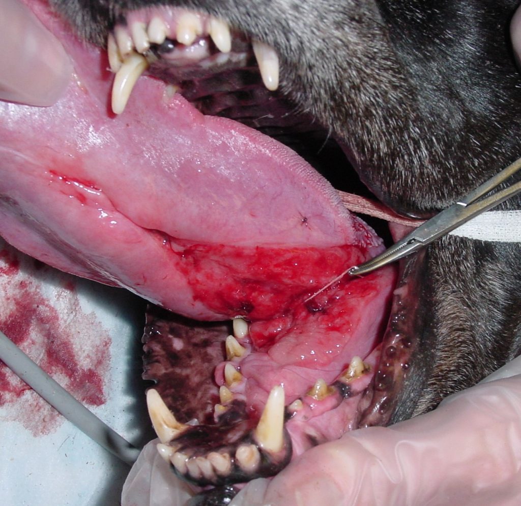
A linear foreign body caught around the lingual frenulum can cause marked swelling and redness beneath the tongue, as well as swelling of the tongue itself. Young cats are the more typical patients, but dogs can be affected. In one unusual case, a 10-year-old dog named Lily had the inner twine of a golf ball wrapped around the base of her tongue.
Chronic irritation can produce another lookalike: a large area of granulation tissue beneath the tongue. A rough tooth surface caused by tooth resorption is one possible source. For clinicians assessing feline sublingual lesions, the practical takeaway is to keep the differential broad and use biopsy when the examination cannot distinguish among these causes.
John Lewis, VMD, DAVDC, FF-OMFS, practices veterinary dentistry and oral surgery at Veterinary Dentistry Specialists and teaches at Silo Academy Education Center, both located in Chadds Ford, Pa.
References
- DeYoung DW, Kealy JK, Kluge IP. Attempts to produce salivary cysts in the dog. Am J Vet Res. 1978;39:185-186.
- Lewis, JR. What you need to know about oral eosinophilic diseases in dogs and cats. July 20, 2015. https://www.veterinarypracticenews.com/what-you-need-to-know-about-oral-eosinophilic-diseases-in-dogs-and-cats/
Discussion
Sign in to join the discussion.