Obstructive urolithiasis poses serious health risks for small ruminants, making early detection and proper management vital for their well-being.
**Understanding Obstructive Urolithiasis in Small Ruminants**
Obstructive urolithiasis is an urgent medical issue frequently seen in small ruminants, particularly with the growing trend of these animals as companion pets. This condition arises from urinary blockages that not only pose immediate risks but can also result in long-term complications like electrolyte imbalances and potential kidney damage. Prompt identification and treatment are essential to improving outcomes for affected animals.
**Critical Signs and Symptoms**
Recognizing the signs of obstructive urolithiasis is crucial for timely intervention. Key indicators include behavioral changes such as straining to urinate or exhibiting discomfort, along with physical symptoms like a tense abdomen or lethargy. In some cases, animals may show distress through tail flagging or vocalizations. An animal that has exhibited symptoms for less than 24 hours typically has a better prognosis; thus, quickly establishing the timeline of clinical signs can significantly impact treatment opportunities.
**Risk Factors for Development**
Several factors contribute to the risk of developing urolithiasis. The anatomy of small ruminants, particularly males, predisposes them due to longer urethras and narrower passages, which can complicate urine flow and create blockages. Diet plays a significant role as well. For example, high-calcium feeds are linked to certain types of uroliths forming in the urinary tract, while alkalinity of urine—often resulting from a forage-heavy diet—can lead to the creation of specific crystals like struvite.
Water intake is another critical factor. When ruminants don't drink enough, their urine becomes concentrated, increasing the likelihood of urolith formation. It's also important to note that while some breeds, such as Nigerian Dwarfs and Pygmy goats, might show higher incidence rates, any small ruminant can be affected regardless of noted risk factors.
**Diagnostic Imaging Techniques**
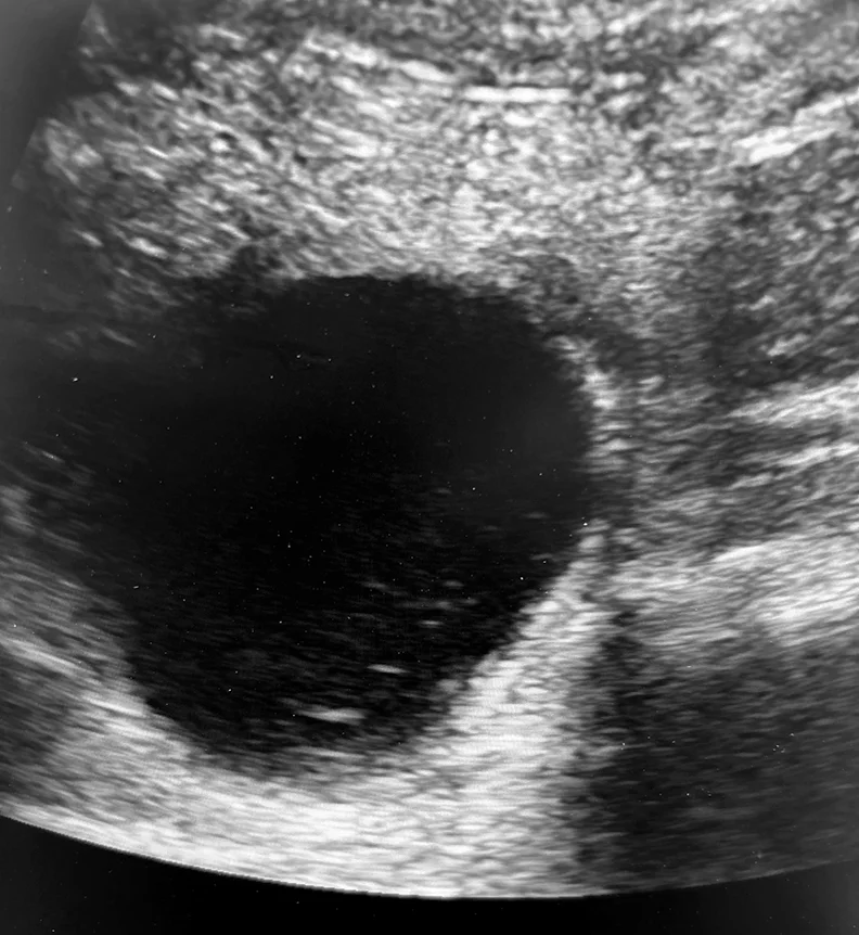
For effective diagnosis, ultrasonography stands out as the go-to method for confirming urinary obstruction. Signs on ultrasound, like a enlarged urinary bladder, are indicative of blockage and warrant immediate medical attention. Traditional radiography can help in identifying the type of urolith but may not always provide clear results, especially in larger animals. Lower diagnostic effectiveness in certain cases means that thorough examination and follow-up are imperative.
In conclusion, understanding the nuances of obstructive urolithiasis, including its signs, risk factors, and diagnostic pathways, is essential for those working with small ruminants. Being alert to early symptoms and recognizing potential pitfalls can lead to more favorable outcomes in these often overlooked but critical situations.
Discussion
Sign in to join the discussion.